Clinical scenario 4: Jasper
Jasper, a two year old boy with asthma, presents to the emergency department with a one week history of upper respiratory tract infection symptoms, followed by increasing coughing and wheezing over the past two days. His mother has been giving him regular salbutamol.
His vital signs are: RR 40/min, HR 150/min, Temp 39ºC and SaO2 90% in room air.
Bronchial breath sounds are present in the right lower zone, with reduced air entry and occasional crackles. There is mild dullness to percussion over the right lower zone.
The following five questions refer to Jasper's clinical scenario.
|
Question 20 Which one of the following is your first step in management? |
|---|
| Nebulised salbutamol |
| Salbutamol via spacer |
| Nebulised adrenaline |
| Prednisolone |
| Oxygen via mask |
| Check answer |
ExplanationThis child is de-saturating and requires oxygen. |
|
Question 21 Which one of the following is the most likely diagnosis? |
|---|
| Asthma |
| Inhaled foreign body |
| Pneumonia |
| Cardiac failure |
| Bronchiolitis |
| Check answer |
ExplanationThe focal nature of the findings are much more likely in pneumonia. Asthma is a possibility but not supported by the findings on auscultation, as well as the fact he has not been responding to salbutamol at home. He is too old for bronchiolitis. A child with cardiac failure would initially have bibasal crackles and generally not be febrile. There are no other features suggesting a cardiac cause, such as oedema, hepatomegaly or a murmur. Inhaled foreign bodies should be considered in a child with recurrent pneumonia or if there was a history of a choking episode. The right main bronchus is the main site for an inhaled foreign body. The preceding history of an URTI makes this less likely. |
|
Question 22 Which one of the following is the most likely organism? |
|---|
| Mycoplasma |
| Human metapneumovirus |
| Staph aureus |
| RSV |
| Streptococcal pneumonia |
| Check answer |
ExplanationStreptococcal pneumonia is the most common cause of bacterial pneumonia at any age. It may be preceded by a mild upper respiratory tract infection, followed by fever, tachypnoea, cough and pleuritic chest pain. Signs may include respiratory distress, dullness to percussion, reduced breath sounds and bronchial breathing over the area involved. Staphylococcal pneumonia usually causes a more severe form of pneumonia with toxicity and more rapid deterioration. Abscesses and marked pleural effusions are more common. Mycoplasma is a frequent cause of pneumonia in children, although it is uncommon in infancy. Features tend to be more non-specific and protracted. RSV and HMPV are common causes of bronchiolitis or viral pneumonia, which is most common under five years of age. They cause all manner of respiratory illnesses. Pneumonia is typically more widespread rather than lobar. |
Jasper's clinical scenario continued
Chest x-ray reveals collapse and consolidation in the right lower lobe. Jasper’s WCC is 21, neutrophils 18 and CRP 140.
|
Question 23 What treatment will you commence? |
|---|
| PO Augmentin |
| PO roxithromycin |
| IV benzylpenicillin |
| IV flucloxacillin |
| IV ceftriaxone |
| Check answer |
ExplanationPenicillin is the first line, unless you suspect a resistant organism or have an organism identified from blood cultures with resistance to penicillin. |
Jasper's clinical scenario continued
Jasper has been managed with intravenous benzylpenicillin for the last two days. He remains febrile to 39ºC, tachycardic, tachypnoeic and lethargic. He has poor air entry and stony dullness to percussion in his right base. He now needs 10L oxygen by non-rebreather mask to maintain saturations of 95%.
|
Question 24 Which one of the following is the best course of action? |
|---|
| Increase the oxygen flow until the oxygen saturations are >95% |
| Increase the penicillin dosage and frequency to 60mg/kg/day |
| Increase the paracetamol dosage and frequency to maximum |
| Repeat the chest x-ray to exclude effusions |
| Continue current treatment as pneumonia requires 7 days therapy |
| Check answer |
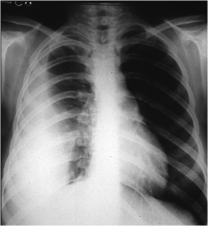
ExplanationClinically, this child has an effusion. CXR will confirm the diagnosis. Depending on the size, this child may need chest drainage. Clinically, that appears likely. Here is a supine chest x-ray with a right-sided pleural effusion:
|
